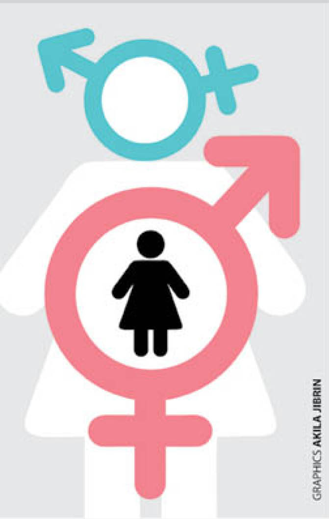
Hermaphroditism: Causes of undifferentiated genitals
What to know about the condition We see 3 hermaphrodites every week – AKTH paediatric surgeon The discovery of seven persons with sex differentiation disorder also known as intersex or hermaphroditism in Kano recently was greeted with awe because of the low awareness about the condition. Professor Mohammad Aminu Mohammad of the Paediatric Surgery […]

Hermaphroditism
-
What to know about the condition
-
We see 3 hermaphrodites every week – AKTH paediatric surgeon
The discovery of seven persons with sex differentiation disorder also known as intersex or hermaphroditism in Kano recently was greeted with awe because of the low awareness about the condition.
Professor Mohammad Aminu Mohammad of the Paediatric Surgery Department, Aminu Kano Teaching Hospital (AKTH), told Daily Trust that every week three patients with cases of ambiguous genitalia are seen in the hospital.
Prof Mohammad said an intersex or a hermaphrodite was a disorder of sex differentiation where patients, children or newborns were born with neither classically female external genitalia nor classical male external genitalia.
He said, “The male external genitalia we know has a penis and two testicles in the scrotum, while female external genitalia has what we call the vestibule that has the urethral opening where the person passes urine and the vaginal opening – that is the genital part of the female, and then the separate anal opening.
“Some people are born with ambiguity; that is, it is neither classically defined as that of a male or classically looking like that of a female.”
He said it was also a social emergency, explaining that the first thing people asked when a child was born was if it was a male or a female.
He explained that, “Sometimes without even asking if the mother or newborn is safe, people want to know what the sex of the child is. If that cannot be determined at that time, then it is a big issue.”
He said while it was a condition that could be remedied, because of lack of knowledge people sometimes did not seek help on time.
“We see this kind of cases at different stages of life. Some come to the hospital early while others live with it until even puberty; when the characteristics of maybe a female gender has started growing in the baby that was hitherto raised as a male or failure of the female external genitalia at puberty to develop,” he said.
Causes of the disorder
According to Prof Mohammad, some of the causes are right from the time of fertilisation while others are as a result of some maternal effects of hormones or in some others as a result of effect of some drugs that are given to the parents.
He said genetic gender was determined at the time of fertilization; whether it was a male developing sperm that has the Y chromosome or it was a female developing sperm that carried the X chromosome.
“So if any one of these fertilises an ovum, it will either lead to the male or a female gender. But in some situations you find out that an empty shell that doesn’t contain X or Y will fertilise the ovum which leads to the development of one category of the abnormality, “he explained.
Explaining how an empty sperm fertilises an ovum, he said, “The sperm is one of the rapidly dividing cells in the body. So sometimes it is one cell that divides into two, to half chromosome of a normal cell and half of the other.
“Sometimes in the process of that division, the whole of the genetic component of that cell to be developed into sperm will go into Y and the other is left empty. So that is the reason why such patients will neither carry the male; that is the Y chromosome, or a female developing X chromosome in it.”
He said there were also cases where you found two sperms fertilising an ovum thereby leading to the abnormality.
The expert further said sometimes a normally fertilised male or a female with either X or Y chromosome would be exposed to some chemicals in the mother; probably because the mother is producing them in excess.
“For example, a mother that is pregnant with a female baby but her adrenal gland produces more testosterone that will stimulate the genital tract to start developing towards a male external genitalia but it will not be complete because there is no genetic stimulation to it.
“And in some patients, it might be because of the treatment the mother is receiving for some conditions that have issues and cause the baby problems developing external genitalia.
“Some of them can also develop when the patient’s adrenal gland is enlarged and producing more testosterone that will be stimulating a normally developing female genitalia to start acquiring some of the characteristics of the male external genitalia.
“This type is what we call the congenital adrenal hyperplexia and is the most common (about 8 to 8.5 out of 10) of all ambiguous genitalia.”
According to the expert, there is also the female pseudo hermaphrodite and male pseudo hermaphrodite.
He explained that, “Sometimes it is the developing testes of a newborn male baby that will not be producing enough testosterone and that will make their external genitalia to look like that of a female. So in the long run, the patient will now acquire one of the characteristics.”
He added that there were four major types of ambiguity in genitalia.
“The female pseudo hermaphrodite, which is the commonest, caused by adrenal congenital hyperplexia, then the male pseudo hermaphrodite – the patient with different genetic composition either 45 XX, 46XY or 47 XY or 47 YYX.
“All of them can now develop to have some abnormalities, these are the true intersex.
“Then the patients who were fertilised by an empty sperm that doesn’t carry X or Y will have what we call digenetic gonad; they don’t have classical testes or classical ovary.
“They will have a mix-up tissue within the gonad, that’s why we don’t call it testes or ovary but gonad.”
On the ones caused by drugs, he said it did not mean they abused drugs, but that sometimes some mothers did not even know that they were pregnant, especially in the early stages, and the drugs administered caused it.
Prevalence in Kano
The surgeon said it was common in Kano with about one to three out of 5,000 live births, explaining that every week, medical experts at the Aminu Kano Teaching Hospital (AKTH) see at least two to three cases.
He, however, added that the hospital was a referral centre for most of the North West states.
He said the major problem was that a lot of the cases were either not recognised were not presented to medical experts.
What is done to determine the sex of the patient?
He explained that the history was first taken to find out what happened from the time the baby was born; where the child was delivered, who assigned the gender, etc.
He said sometimes medical experts found that it was lack of thoroughly looking to determine the sex, and that the approach towards determining the sex also depended on the age of the patient, adding that some patients came when they had already developed beards, breasts or reached puberty.
He further said the next step was the physical examination of the patient.
“We all know that a typical male external genitalia has a normally developed penis and two testicles while the female patient will not have testes, will not have enlarged penis, the clitoris will be small, then there are two openings in the perineum, apart from the anus, the urethra and the vaginal opening.
“So in this condition we examine thoroughly under good light to see if the patient is being labelled a male while she is actually a female, or being labelled a female while actually a male.”
Prof Mohammad said there were some that could not be differentiated externally even with physical examination, hence medical experts did genetic analysis to know whether the patient’s genetic composition was “46XX, which is for a female or 46XY which is for a male, and sometimes we see some patients that have half of their sex as 46XX and the other half 46XY. These are the true intersex.”
The expert said for some patients, ultrasound scan was done to see the internal organs and, “If the testicles are not palpable we look for them around their route of descent, if the ultra sound locates those testicles, we want to explore and examine.
“Also, if ultra sound suggests that there is no uterus, no fallopian tube, no ovaries, but we have seen testes then we are dealing with a male.
“If the ultrasound didn’t detect any of that, then probably we are dealing with a male, but if it shows that there are ovaries, fallopian tube and uterus, then we are dealing with a female child.”
He further said the final step was to do mini-laparotomy, which involved opening the lower abdomen.
“For instance, if there are no uterus, fallopian tube and ovaries, then we are dealing with a male, if these organs are there then we are dealing with a female,” he explained.
He said medical experts looked at the genitalia to see which one was likely to give the child a better function and advised parents to let the child be a male or a female, and that with that, they had to do a lot of reconstruction that would help the patient.
He said, “But there are few cases, thank God, where the internal organs are neither testes nor ovaries. These kinds of patients are the true intersex or the digenetic gonads.”
He added that gonads that contained both ovarian and testicular tissues were often removed because of the risk of becoming cancer in the patient.
